


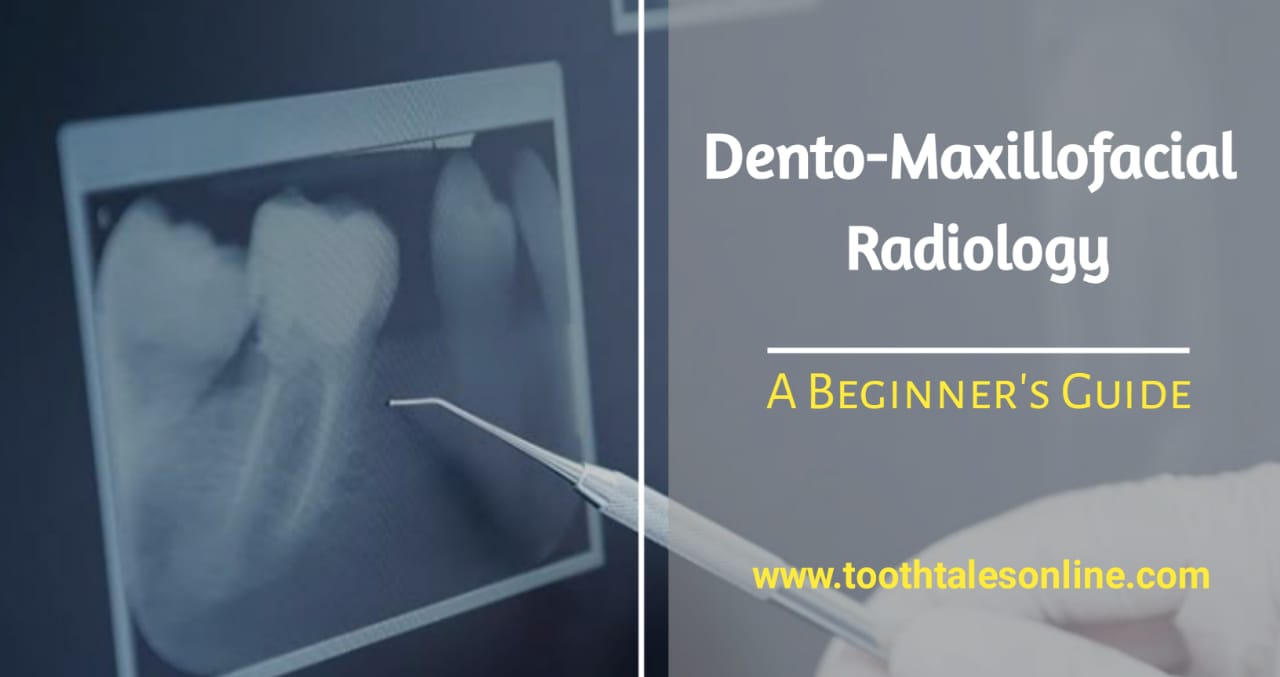
Introduction:
Dento-Maxillofacial Radiology might sound like a mouthful, but it’s actually a crucial part of dental and medical care. Simply put, it’s a special type of X-ray imaging that helps dentists and doctors see inside your mouth, jaw, and face. Think of it as taking a detailed picture with a machine that uses X-rays instead of regular light. Let’s break down what dento-maxillofacial radiology is, the different types of dental X-rays, why it matters, and how it keeps you safe.
What is Dento-Maxillofacial Radiology?
Dento-Maxillofacial Radiology is a branch of medical imaging focused on capturing detailed images of your teeth, jaws, face, and surrounding areas using various X-ray techniques. These images, known as radiographs or X-rays, provide essential information to dentists, oral surgeons, and other healthcare professionals. They help in diagnosing and treating a wide range of dental and facial conditions.
Key Points:
- Medical Imaging: Uses X-rays to see inside the mouth and face.
- Diagnostic Tool: Helps identify dental issues like cavities, bone loss, and tooth alignment problems.
- Collaborative Use: Utilized by dentists, oral surgeons, orthodontists, and other specialists.
Types of Dento-Maxillofacial Radiographs:
There are several types of dental X-rays, each serving a specific purpose. Understanding these can help you know what to expect during your dental visits.
1. Intraoral Radiographs
Intraoral radiographs are the most common type of dental X-rays. They involve placing a sensor or film inside your mouth to capture detailed images of individual teeth, roots, and the surrounding bone structure.
Types of Intraoral Radiographs:
- Bitewing X-rays: Show the upper and lower teeth in one area of the mouth. They help detect cavities between teeth and assess bone levels.
- Periapical X-rays: Capture the entire tooth from the crown to the root, including the surrounding bone. These are useful for diagnosing issues like abscesses or impacted teeth.
- Occlusal X-rays: Larger than other intraoral X-rays, they show the floor of the mouth and help detect extra teeth, impacted teeth, or jaw fractures.
2. Extraoral Radiographs
Extraoral radiographs capture images from outside the mouth. They provide a broader view of the jaw, skull, and facial structures.
Types of Extraoral Radiographs:
- Panoramic Radiograph: Offers a wide view of all teeth, upper and lower jaws, and surrounding structures. It’s particularly useful for assessing wisdom teeth, jaw fractures, and overall dental development.
- Cephalometric Radiograph: Primarily used in orthodontics, this side-view image of the head helps evaluate the relationship between teeth, jaws, and facial structures. Orthodontists use it to plan braces and other treatments.
- Cone Beam Computed Tomography (CBCT): Produces detailed 3D images of teeth, jaws, and surrounding tissues. CBCT is valuable for complex procedures like dental implant placement, orthognathic surgery, and diagnosing temporomandibular joint (TMJ) disorders.
Why Dento-Maxillofacial Radiology Matters?
Dento-Maxillofacial Radiology is essential in modern dental and medical practices for several reasons. It enhances the ability to diagnose, plan treatments, monitor health, improve patient care, and support research and education.
1. Accurate Diagnosis
Detailed X-ray images help dentists and doctors accurately diagnose various conditions, such as:
- Dental Caries (Cavities): Detect early signs of tooth decay that might not be visible during a regular dental exam.
- Periodontal Disease: Assess bone loss around teeth, which is crucial for treating gum disease.
- Oral Infections: Identify abscesses or other infections within the mouth and jaw.
- Tooth Development Abnormalities: Spot issues with tooth alignment or eruption.
2. Treatment Planning
Radiographic images are vital for planning various dental treatments, including:
- Tooth Extractions: Understand the position of roots and surrounding bone to safely remove teeth.
- Root Canal Therapy: Visualize the tooth’s root structure to effectively perform root canals.
- Dental Implant Placement: Ensure accurate placement of implants by assessing bone density and structure.
- Orthodontic Treatment: Plan braces or aligners by analyzing jaw and tooth relationships.
- Oral Surgeries: Prepare for surgeries by mapping out the area in detail.
3. Monitoring Dental Health
Regular dental X-rays allow dentists to monitor changes in your oral health over time. They help track:
- Progression of Dental Conditions: See how cavities or gum disease are developing.
- Effectiveness of Treatments: Evaluate if treatments like fillings or implants are working as intended.
- Early Detection of Problems: Identify potential issues before they become serious, such as early signs of oral cancer.
4. Improving Patient Care
By providing detailed diagnostic information, dento-maxillofacial radiology contributes to better patient outcomes. It allows:
- Personalized Treatments: Tailor treatments to each patient’s unique needs and conditions.
- Minimized Risks: Anticipate and avoid potential complications during procedures.
- Enhanced Success Rates: Increase the likelihood of successful treatments through precise planning.
5. Research and Education
Dento-maxillofacial radiology is also crucial for advancing dental and medical knowledge. It supports:
- Research: Helps scientists study oral diseases, treatment outcomes, and develop new techniques.
- Education: Trains future dentists, oral surgeons, and radiologists to interpret X-rays accurately and make informed clinical decisions.
Safety Precautions in Dento-Maxillofacial Radiology:
While dental X-rays are incredibly useful, it’s essential to ensure patient safety by following strict radiation protection guidelines. Here are some key safety measures:
1. Use of Lead Aprons and Thyroid Collars:
To minimize radiation exposure, patients should wear lead aprons and thyroid collars during X-ray imaging. These protective garments shield vital organs from unnecessary radiation.
2. Limiting Radiation Exposure:
Dentists and radiology technicians aim to use the lowest possible radiation dose needed to obtain clear images. Advances in digital radiography have significantly reduced radiation exposure compared to traditional film-based X-rays.
3. Proper Technique and Positioning:
Correctly positioning the X-ray machine and the patient’s head is crucial for obtaining accurate images while minimizing radiation exposure to non-target areas. Proper technique ensures high-quality images with minimal radiation.
4. Pregnancy Precautions:
Pregnant patients should inform their dentist or healthcare provider before undergoing any radiographic procedures. Although dental X-rays emit low levels of radiation, extra precautions are taken to protect the developing fetus, such as using lead aprons and avoiding unnecessary X-rays.
How Dento-Maxillofacial Radiology Enhances Your Dental Experience?
Understanding the role of dento-maxillofacial radiology can help you appreciate its importance in your dental care. Here’s how it enhances your overall dental experience:
Early Detection and Prevention:
Regular dental X-rays can catch problems early, allowing for timely intervention. Early detection often means simpler and less invasive treatments, saving you time, money, and discomfort.
Comprehensive Treatment Plans:
With detailed images, your dentist can create comprehensive treatment plans that address all aspects of your oral health. Whether you need a filling, orthodontic treatment, or a dental implant, accurate imaging ensures the best possible outcomes.
Personalized Care:
Every mouth is unique, and dento-maxillofacial radiology allows for personalized care tailored to your specific needs. From understanding your bone structure to planning cosmetic dental procedures, detailed images help your dentist provide customized treatments.
Enhanced Communication:
Clear radiographic images improve communication between you and your dental care team. They help you understand your dental issues and the proposed treatments, making it easier to make informed decisions about your oral health.
Advancements in Dento-Maxillofacial Radiology:
The field of dento-maxillofacial radiology is continually evolving, incorporating new technologies to improve diagnostic capabilities and patient safety.
Digital Radiography:
Digital X-rays have revolutionized dental imaging by providing instant images with higher resolution and lower radiation doses compared to traditional film-based X-rays. They also allow for easy storage, sharing, and manipulation of images for better diagnosis and treatment planning.
Cone Beam Computed Tomography (CBCT):
CBCT offers 3D imaging, providing a more detailed view of the teeth, jaws, and surrounding structures. This technology is especially useful for complex cases, such as:
- Dental Implants: Ensuring accurate placement by assessing bone quality and quantity.
- Orthognathic Surgery: Planning corrective jaw surgeries with precise measurements.
- TMJ Disorders: Diagnosing issues with the temporomandibular joint with greater accuracy.
Artificial Intelligence (AI) in Radiology:
AI is being integrated into dento-maxillofacial radiology to assist in image analysis. AI algorithms can help detect abnormalities, measure bone density, and even predict treatment outcomes, enhancing the accuracy and efficiency of diagnoses.
Common Myths About Dental X-Rays:
There are several misconceptions about dental X-rays that can cause unnecessary worry. Let’s debunk some common myths:
Myth 1: Dental X-Rays Are Dangerous
Fact: Dental X-rays use very low levels of radiation, much lower than many other everyday sources. Modern digital X-rays further reduce exposure, making them safe when used appropriately.
Myth 2: You Can’t Have an X-Ray Without a Problem
Fact: Regular dental X-rays are essential for maintaining oral health. They help detect issues before symptoms appear, allowing for early and effective treatment.
Myth 3: Dental X-Rays Are Painful
Fact: Dental X-rays are quick and painless. The process involves placing a small sensor in your mouth and taking the image, which only takes a few seconds.
Myth 4: X-Rays Can Cause Cancer
Fact: The radiation exposure from dental X-rays is minimal and not linked to cancer when used correctly. Dentists follow strict guidelines to ensure patient safety.
Conclusion:
Dento-Maxillofacial Radiology is a vital part of dental and medical care, providing detailed images that help diagnose and treat a wide range of oral and facial conditions. From detecting cavities and gum disease to planning complex surgeries and orthodontic treatments, dental X-rays play a crucial role in maintaining your oral health. By following strict safety protocols and embracing technological advancements, dento-maxillofacial radiology ensures that you receive the best possible care with minimal risks. Understanding this field can help you appreciate the importance of regular dental X-rays and the role they play in keeping your smile healthy and bright.
Frequently Asked Questions (FAQs):
To help you better understand dento-maxillofacial radiology, here are some of the most commonly asked questions:
1. How Often Do I Need Dental X-Rays?
The frequency of dental X-rays depends on your individual oral health needs. Generally:
- Routine Checkups: Every 1-2 years for healthy individuals.
- High-Risk Patients: More frequent X-rays if you have a history of dental issues or are at higher risk for cavities and gum disease.
- Specific Treatments: As needed for procedures like implants or orthodontics.
2. Is It Safe to Have Dental X-Rays?
Yes, dental X-rays are safe when performed by trained professionals. They use low levels of radiation, and safety measures like lead aprons and digital technology further reduce exposure.
3. What Should I Do If I’m Pregnant and Need a Dental X-Ray?
If you’re pregnant, inform your dentist before any X-rays. They will take precautions to minimize radiation exposure, such as using a lead apron and only performing necessary X-rays.
4. How Do Digital X-Rays Compare to Traditional Film X-Rays?
Digital X-rays are faster, use less radiation, and provide higher-resolution images compared to traditional film X-rays. They also allow for easy storage and sharing of images.
5. Can I See My Dental X-Rays?
Absolutely! Your dentist can show you your X-ray images and explain what they reveal about your oral health. It’s a great way to understand your dental condition and treatment options.
6. What Happens During a Panoramic X-Ray?
During a panoramic X-ray, you’ll sit or stand still while the machine moves around your head, capturing a broad view of your teeth, jaws, and surrounding structures in one image. It’s quick and painless.
7. Are There Any Risks Associated with CBCT Scans?
CBCT scans provide detailed 3D images with higher radiation doses than standard X-rays. However, they are only recommended when necessary for complex cases. Your dentist will weigh the benefits against the risks before ordering a CBCT scan.
8. How Do I Prepare for a Dental X-Ray?
Generally, no special preparation is needed. Inform your dentist if you’re pregnant or have any concerns. For certain types of X-rays, you might need to remove any metal objects or bite down on a sensor.
9. Can Children Have Dental X-Rays?
Yes, children can have dental X-rays. Dentists use child-friendly techniques and minimal radiation doses to ensure safety while effectively diagnosing dental issues.
10. What Are the Signs That I Might Need a Dental X-Ray?
Signs you might need a dental X-ray include:
- Persistent tooth pain or sensitivity
- Swelling or gum irritation
- Visible cavities or tooth decay
- Planning for orthodontic treatment or implants
- Assessing overall oral health during regular checkups


Add Comment